Cell division protein

From Proteopedia

Jump to: navigation, search

Contents

Bacillus subtilis division protein

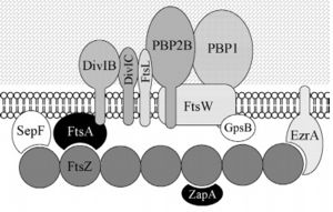
Bacillus subtilis is a prokaryotic organism, a Gram-positive bacterium, and thus has a cytoplasmic membrane plus a thick cell wall made of peptidoglycan and associated anionic polymers, such as teicoic acid. Bacteria are recognized for the reproductive success and conquest of various environments on Earth, with the presence of a complex and sophisticated machinery of cell division and formation of identical daughter cells a potent factor in this success. There are currently 24 proteins known to be associated with division in B. subtilis: ClpX, DivIB,DivIC, DivIVA, EzrA, FtsA, FtsL, FtsW, FtsZ,GpsB, MciZ, MinC, MinD, MinJ, Noc, PBP1,PBP2B, SepF, SftA, SpoIIE, SpoIIIE, UgtP, YneA and ZapA. These proteins can be divided in two main groups: proteins that make up the divisome - macromolecular complex composed of about 20 proteins, which promotes the construction of the cell wall and cytoplasmic membrane, forming the division septum (DivIB, DivIC, EzrA, FtsA, FtsL, FtsW,FtsZ, GpsB, PBP1, PBP2B, SepF and ZapA) and proteins that regulate the assembly of the divisome (ClpX, DivIVA, MciZ, MinC, MinD, MinJ,Noc, UgtP and YneA).

B. subtilis divisome
Among these, the FtsZ stands out for acting in the recruitment of other proteins for the formation of the constriction ring that culminates in cell division.
  • FtsN is essential in activating septal peptidoglycan synthesis and constriction of the cell.
  • FtsQ is essential in for early and late bacterial cell wall division[1].
  • FtsE and FtsX plays a central role by recruiting proteins to the divisive apparatus in bacteria [2]
  • FtsY is a bacterial single-recognition particle receptor [3]
  • ZapA, ZapB, ZapC and ZapD are recruited by FtsZ into the Z-ring forming during cell wall division[4],[5].
  • ZipA links FtsZ to the cytoplasmic membrane[6].
  • SepF align FtsZ filaments in cyanobacteria[7].
  • DedA is part of the constriction phase of the bacterial cell division cycle[8].

General Function

FtsZ (Filamentation temperature sensitive Z) is the main coordinator of septum formation and the most widely conserved division protein, being present in essentially all bacterial genomes that have been sequenced to date[9]. In eubacteria, the FtsZ gene is usually found in the dcw gene cluster, a DNA region containing division and cell-wall synthesis gene. This protein is extremely important to binary fission - more specifically, formation of the Z ring - in rod-shaped bacteria entails the formation of a transverse septum that divides a progenitor cell into two equal-sized daughter cells. Septum formation is faithfully coordinated with chromosome replication and segregation and the spatial control, on the other hand, is evident in the placement of the newly formed septum at precisely the middle of the progenitor cell, which ensures that the two daughter cells generated are morphologically and genetically equivalent. It means that despite the apparent simplicity, cell division in bacteria is subject to tight spatiotemporal control.

Structure and Biochemistry of FtsZ

FtsZ is a tubulin-like protein, which is widely conserved in bacteria and the main component of the bacterial cytokinesis machine, or “divisome.” FtsZ is a 40 kDa protein that folds into two independent globular domains [N-terminal (1-203) and C-terminal (204-316)] and has an unstructured tail of about 50 amino acids followed by a 15–17 conserved amino acid sequence at its extreme C-terminus. This conserved terminal sequence is also known as the ‘C-terminal peptide’ (CTP), since it is in the N-terminal domain that the nucleotide binding region is contained. Self-assembly of FtsZ involves interactions between the C-terminal globular domain of one subunit with the N-terminal globular domain of another subunit. The CTP, on the other hand, is the binding site for several of the proteins that interact with FtsZ. The N-terminal and C-terminal domain are separated by the central H7 helix (178-202). FtsZ and tubulin share several essential properties: their assembly is cooperative, stimulated by GTP, and leads to GTP hydrolysis; they form dynamic polymers whose turnover is dependent on nucleotide hydrolysis; they use essentially the same bond for polymer formation; and recent evidence indicates that they undergo similar allosteric transitions upon polymerization. The folding of the FtsZ N-terminal domain is typical of GTPases, with six parallel β-strands (S1-S6) surrounded by six α-helices (H1-H6), named according to the tubulin structure (show secondary structure). The C-terminal domain is formed by four parallel β-strands (S7-S10) surrounded by two helices, with the antiparallel strand S10. The residues in the T1-T4 loops make contact with the phosphate groups of the GDP. The T5 loop between S5 and H5 helix contains residues that make hydrogen bonds with the sugar moiety and also contacts with the phosphate of GDP, while interactions with the nucleotide nitrogen base are done by residues of the H7 helix.

Polymerization mechanism

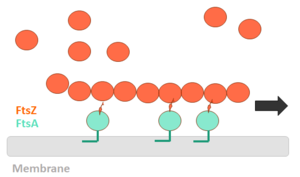
In order for polymerization of the monomers of FtsZ to occur, there is a need for interaction of the N-terminal nucleotide binding domain with the C-terminal domain of another monomer. The formation of a complete GTP hydrolysis site depends on the positioning of acid residues of the T7 loop at the nucleotide binding site of the prior monomer in the polymer, explaining why the GTPase activity of FtsZ only occurs when the protein is in the polymer form. The GTP binding site (PDB code 1w5b). Water molecules are shown as red spheres. The fact that FtsZ GTPase activity interferes with the dynamics of its autoassociation and the morphology of its polymers, as well as with microtubules and actin, opens the possibility that FtsZ can also present dynamic treadmilling or instability. Dynamic FtsZ beams were observed in vitro and protofilaments exhibiting treadmilling dependent on FtsA protein.

FtsZ polimerization mechanism

The binding of GTP promotes the longitudinal association of the monomers forming protofilaments, and the hydrolysis of the nucleotide leads to depolymerization and consequent disorganization of the protofilament. FtsZ protofilaments have a strong tendency to associate further to form multi-stranded polymers. The tendency of FtsZ filaments to form lateral interactions in greatly increased by the presence of cations such the Ca2+. However, there is still controversy regarding the mechanism of polymerization of FtsZ and details of the process are being elucidated. What is currently in place is that loop motion between the H6-H7 helices away from the nucleotide cavity (downward movement of the H7 helix, and 23-degree rotation of the C-terminal domain relative to the N-terminal), creates a groove between the C-terminal domain and the H7 helix that does not exist in the structures described above. This conformation promotes the insertion of the T7 loop in the active site of another monomer, which in the presence of divalent cation would stabilize a dimeric interface with more extensive contacts leading to polymerization. Therefore, the "assembly switch" would be the conversion of a monomer into the closed conformation (without the groove between H7 and C-terminal) to the open conformation (with the groove between H7 and the C-terminus), the latter competent for polymerization.

Relevance

The understanding of the structure of FtsZ and its polymerization process is relevant because it can be used as a target for the design of new antibiotics capable of selectively combating bacterial infections. This is due to its structural and functional conservation in prokaryotes, to their essential role in the replication of these organisms, and the evolutionarily distant from tubulin, its counterpart in eukaryotes. Thus, the use of an antibiotic would not cause effects on eukaryotic tubulin.

3D structures of cell division protein

Cell division protein 3D structures

FtsZ of Bacillus subtilis complex with sulfate (PDB code 2vam)

Drag the structure with the mouse to rotate

References

[10] [11] [12] [13] [14] [15] [16] [17]

  1. van den Ent F, Vinkenvleugel TM, Ind A, West P, Veprintsev D, Nanninga N, den Blaauwen T, Lowe J. Structural and mutational analysis of the cell division protein FtsQ. Mol Microbiol. 2008 Apr;68(1):110-23. Epub 2008 Feb 26. PMID:18312270 doi:http://dx.doi.org/10.1111/j.1365-2958.2008.06141.x
  2. Alcorlo M, Straume D, Lutkenhaus J, Havarstein LS, Hermoso JA. Structural Characterization of the Essential Cell Division Protein FtsE and Its Interaction with FtsX in Streptococcus pneumoniae. mBio. 2020 Sep 1;11(5). pii: mBio.01488-20. doi: 10.1128/mBio.01488-20. PMID:32873757 doi:http://dx.doi.org/10.1128/mBio.01488-20
  3. Angelini S, Deitermann S, Koch HG. FtsY, the bacterial signal-recognition particle receptor, interacts functionally and physically with the SecYEG translocon. EMBO Rep. 2005 May;6(5):476-81. PMID:15815684 doi:10.1038/sj.embor.7400385
  4. Galli E, Gerdes K. Spatial resolution of two bacterial cell division proteins: ZapA recruits ZapB to the inner face of the Z-ring. Mol Microbiol. 2010 Jun;76(6):1514-26. PMID:20487275 doi:10.1111/j.1365-2958.2010.07183.x
  5. Durand-Heredia J, Rivkin E, Fan G, Morales J, Janakiraman A. Identification of ZapD as a cell division factor that promotes the assembly of FtsZ in Escherichia coli. J Bacteriol. 2012 Jun;194(12):3189-98. doi: 10.1128/JB.00176-12. Epub 2012 Apr, 13. PMID:22505682 doi:http://dx.doi.org/10.1128/JB.00176-12
  6. Mosyak L, Zhang Y, Glasfeld E, Haney S, Stahl M, Seehra J, Somers WS. The bacterial cell-division protein ZipA and its interaction with an FtsZ fragment revealed by X-ray crystallography. EMBO J. 2000 Jul 3;19(13):3179-91. PMID:10880432 doi:10.1093/emboj/19.13.3179
  7. Duman R, Ishikawa S, Celik I, Strahl H, Ogasawara N, Troc P, Lowe J, Hamoen LW. Structural and genetic analyses reveal the protein SepF as a new membrane anchor for the Z ring. Proc Natl Acad Sci U S A. 2013 Nov 11. PMID:24218584 doi:http://dx.doi.org/10.1073/pnas.1313978110
  8. Liu B, Hale CA, Persons L, Phillips-Mason PJ, de Boer PAJ. Roles of the DedD Protein in Escherichia coli Cell Constriction. J Bacteriol. 2019 Mar 26;201(8):e00698-18. PMID:30692172 doi:10.1128/JB.00698-18
  9. Silber N, Matos de Opitz CL, Mayer C, Sass P. Cell division protein FtsZ: from structure and mechanism to antibiotic target. Future Microbiol. 2020 Jun;15:801-831. doi: 10.2217/fmb-2019-0348. Epub 2020 Jul , 21. PMID:32692252 doi:http://dx.doi.org/10.2217/fmb-2019-0348
  10. Bisson-Filho AW, Discola KF, Castellen P, Blasios V, Martins A, Sforca ML, Garcia W, Zeri AC, Erickson HP, Dessen A, Gueiros-Filho FJ. FtsZ filament capping by MciZ, a developmental regulator of bacterial division. Proc Natl Acad Sci U S A. 2015 Apr 6. pii: 201414242. PMID:25848052 doi:http://dx.doi.org/10.1073/pnas.1414242112
  11. Wang, X. & Lutkenhaus, J. FtsZ ring: the eubacterial division apparatus conserved in archaebacteria.Mol. Microbiol. 21, 313–319 (1996). Gueiros-Filho, F. J. & Losick, R. A widely conserved bacterial cell division protein that promotes assembly of the tubulin-like protein FtsZ. Genes Dev. 16, 2544–2556 (2002).
  12. Wang, X., Huang, J., Mukherjee, A., Cao, C., and Lutkenhaus, J. (1997). Analysis of the interaction of FtsZ with itself, GTP, and FtsA. J. Bacteriol. 179, 5551–5559.
  13. Vaughan S, Wickstead B, Gull K, Addinall SG. Molecular evolution of FtsZ protein sequences encoded within the genomes of archaea, bacteria, and eukaryota. J Mol Evol. 2004 Jan;58(1):19-29. doi: 10.1007/s00239-003-2523-5. PMID:14743312 doi:http://dx.doi.org/10.1007/s00239-003-2523-5
  14. Szwedziak P, Wang Q, Bharat TA, Tsim M, Lowe J. Architecture of the ring formed by the tubulin homologue FtsZ in bacterial cell division. Elife. 2014 Dec 9;3:e04601. doi: 10.7554/eLife.04601. PMID:25490152 doi:http://dx.doi.org/10.7554/eLife.04601
  15. Huecas, S. et al. Energetics and geometry of FtsZ polymers: nucleated self-assembly of single protofilaments. Biophys. J. 94, 1796–1806 (2008).
  16. Lan, G. et al. (2009) Condensation of FtsZ filaments can drive bacterial cell division. Proc. Natl. Acad. Sci. U. S. A. 106, 121–126
  17. FILHO, Frederico Gueiros. Cell Division. In: GRAUMANN, Peter L. et al. Bacillus: Cellular and Molecular Biology. Germany: Caister Academic Press, 2017.
Personal tools