We apologize for Proteopedia being slow to respond. For the past two years, a new implementation of Proteopedia has been being built. Soon, it will replace this 18-year old system. All existing content will be moved to the new system at a date that will be announced here.
Parvalbumin
From Proteopedia
| |||||||||||
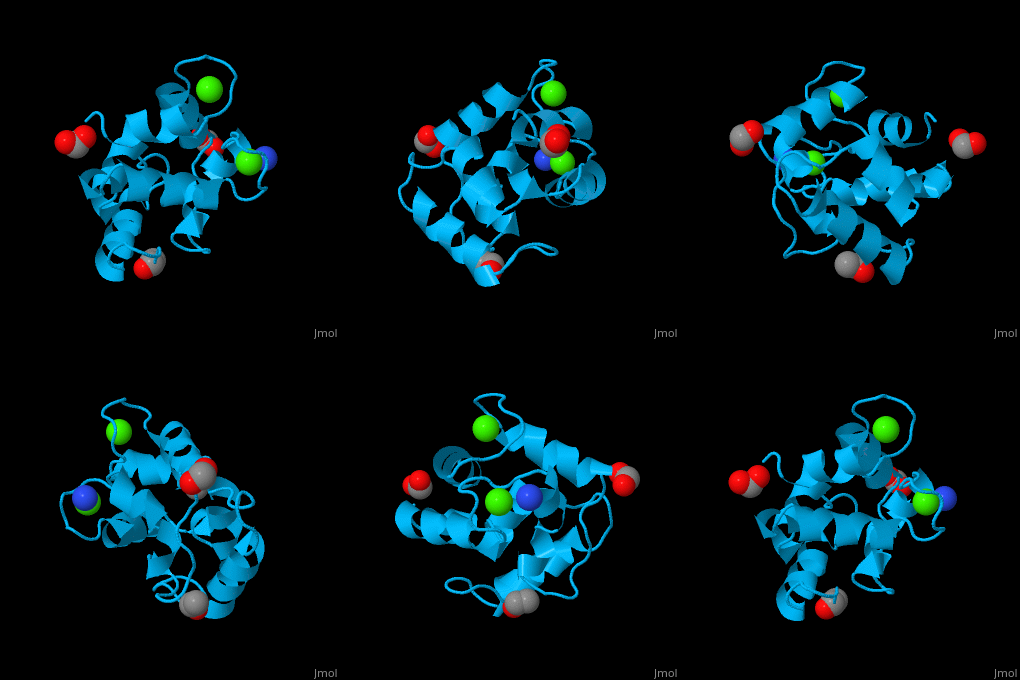
3D Structures of Parvalbumin
Updated on 05-December-2021
References
- ↑ Cates MS, Teodoro ML, Phillips GN Jr. Molecular mechanisms of calcium and magnesium binding to parvalbumin. Biophys J. 2002 Mar;82(3):1133-46. PMID:11867433 doi:http://dx.doi.org/10.1016/S0006-3495(02)75472-6
- ↑ Fohr UG, Weber BR, Muntener M, Staudenmann W, Hughes GJ, Frutiger S, Banville D, Schafer BW, Heizmann CW. Human alpha and beta parvalbumins. Structure and tissue-specific expression. Eur J Biochem. 1993 Aug 1;215(3):719-27. PMID:8354278
- ↑ Lim DL, Neo KH, Yi FC, Chua KY, Goh DL, Shek LP, Giam YC, Van Bever HP, Lee BW. Parvalbumin--the major tropical fish allergen. Pediatr Allergy Immunol. 2008 Aug;19(5):399-407. doi:, 10.1111/j.1399-3038.2007.00674.x. Epub 2008 Jan 25. PMID:18221468 doi:http://dx.doi.org/10.1111/j.1399-3038.2007.00674.x
- ↑ Declercq JP, Evrard C, Lamzin V, Parello J. Crystal structure of the EF-hand parvalbumin at atomic resolution (0.91 A) and at low temperature (100 K). Evidence for conformational multistates within the hydrophobic core. Protein Sci. 1999 Oct;8(10):2194-204. PMID:10548066

