Function
Ribulose-1,5-bisphosphate carboxylase oxygenase – RuBisCO (RBCO) catalyzes the first step in photosynthetic carbon fixation, and it is the most abundant protein on earth. RBCO can either carboxylate or oxygenate ribulose-1,5-bisphosphate (RUBP) with CO2 or O2, respectively. RBCO from flowering plants consists of eight large subunits and eight small subunits.
See Calvin cycle, Rubisco and Crop Output, and RuBisCO (Hebrew).
Structural Features
Quaternery Structure
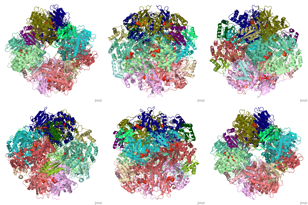
The structure of the Rubisco from spinach is shown in spacefill with its 8 large subunits in shades of blue and and its 8 small subunits in shades of yellow. The large subunits are arranged in head-to-toe pairs like staves in a barrel and the small subunits are arranged at the ends of the barrel. The large subunits contain the active sites and the function of the small subunits is not understood.
Large Subunit Structure
This isolated shows that each subunit has a large C-terminal lobe and a small N-terminal lobe, and the subunits are arranged head-to-toe (antiparallel). are located in the interface of the large subunit pair. The subunits are shown in cartoon with one shown in the secondary structure color scheme. Each active site is occupied by RUBP, which is shown in CPK spacefill. Here is a showing that both lobes contain alpha helices (pink) and beta strands (yellow). The large lobe is dominated by an (amino acids 166-409), which contributes most of the residues that form the active site. One residue from the N-terminal lobe of the adjacent large subunit completes the active site. This scene shows RUBP in spacefill and CPK in one of the active sites in the dimer. Both subunits are shown in transparent cartoon with the α-β barrel is pink and yellow. Asn 123 from the adjacent subunit is in blue spacefill, and residues 121-129 are shown in blue cartoon. This residue does not contribute to catalysis, and it will not be considered further.
Active Site Structure
The structure of spinach Rubisco bound to the naturally occurring inhibitor 2-carboxylarabinitol-1,5-bisphosphate (CAP) and Mg
2+ (
8ruc[1]), implicates residues that are involved in the catalytic mechanism
Image:RubiscoMechanism.pdf.
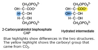
The structure of CAP (left figure) is similar to the hydrated reaction intermediate that is formed following the addition of CO
2 to RUBP. Here is an (cartoon and colored for secondary structure) with CAP and and Mg
2+ in CPK spacefill. This in which the helices have been removed, shows that CAP sits at one end of the α-β barrel, and only residues from the beta strands (gold ball & stick) and loops that link them to helices (white ball & stick) are involved in binding RUBP and Mg
2+ (the RUBP-bidning residue contributed by the N-terminal lobe of the adjacent subunit is not shown). The involved are
acidic residues that interact with Mg
2+,
basic residues and
histidines that interact with phosphate and hydroxyl groups,
polar residues that interact with hydroxyl groups, one
hydrophobic residue, and backbone atoms (white ball & stick) of several residues.
are shown shown here in CPK ball & stick. Asp 203 and Glu 204 bind to and position the magnesium ion. The carbamylated lysine residue KCX 201 coordinates Mg2+ and initiates catalysis by extracting a proton from C3 of RUBP. Note the proximity of the carbamyl group to carbon 3 in this structure. His 294 acts as a catalytic base in the carboxylation step of the mechanism and accepts a proton from the hydroxyl of carbon 3. Mg2+ is coordinated by six ligands. In addition to oxygen atoms in the three residues already mentioned, the ion binds to two oxygen atoms of RUBP. The 6th ligand is either water or in the carboxylation step it binds the incoming CO2. In the structure shown, Mg2+ is bound to the carboxyl group in CAP that corresponds to the fixed CO2 in the hydrated intermediate.
3D structures of RuBisCO
RuBisCO 3D structures


