Introduction
The SecA ATPase SecA or preprotein translocase subunit SecA drives the post-translational translocation of proteins through the SecY channel in the bacterial inner membrane. SecA is a dimer that can dissociate into monomers under certain conditions. Many bacterial proteins are transported post-translationally across the inner membrane by the Sec machinery, which consists of two essential components (1-4). One is the SecY complex, which forms a conserved heterotrimeric protein-conducting channel in the inner membrane.[1] The other is SecA, a cytoplasmic ATPase, which "pushes" substrate polypeptide chains through the SecY channel.[1] . For additional details see SecA PBD motions.
Structure
SecA SecA consists of two RecA-like nucleotide-binding domains (NBD1 and NBD2), which bind the nucleotide between them, a polypeptide-cross-linking domain (PPXD), a helical scaffold domain (HSD) and a helical wing domain (HWD)[2] Although several crystal structures of isolated SecA have been determined, the function of the different domains and the mechanism by which SecA moves polypeptides through the channel remain unknown. Disulphide cross-linking experiments suggest that SecA binds by its NBD1 domain to a non-translocating SecY copy, and moves the polypeptide chain through a neighbouring SecY molecule6. These and other experiments indicate that SecA functions as a monomer during translocation[2]but the issue remains controversial.[2]
Here we report crystal structures of SecA bound in an intermediate state of nucleotide hydrolysis to the SecY channel. The structures suggest mechanisms for how the channel is opened and prepared for the arrival of a translocation substrate, and how SecA moves polypeptides through the channel.
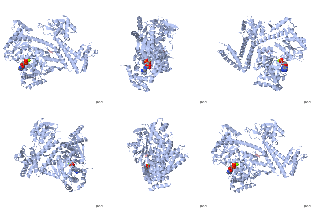
Notable finding is that ADP binding to the high-affinity site stabilizes a compact conformation of SecA (ground state) that has low affinity for the SecYEG/membrane[1]. Click the green link to view the active site of with surrounding amino acids (Water molecules shown as red spheres). This result suggests that following ATP hydrolysis, the ADP-bound SecA undergoes retraction from the translocon to complete one reaction cycle. However, the apo (nucleotide free) form of SecA can also exist in a compact conformation with low affinity for the translocon[1]. Moreover, ADP release from SecA is stimulated by SecYEG/membrane, raising the question whether SecA retraction from the membrane occurs in the ADP-bound form, the apo form, or both[1].
Structure Determination Of SecA-SecY Complexes
SecA Crystallized complexes containing Bacillus subtilis SecA without its non-essential carboxy-terminal domain, and either Thermotoga maritima SecYE or Aquifex aeolicus SecYEG. These crystals diffracted X-rays to a maximum resolution of 6.2 Å and 7.5 Å, respectively. A higher resolution data set (4.5 Å) was obtained for a complex in which both partners were from T. maritima and the SecYEG complex was seleno-methionine (Se-Met) derivatized. All complexes were crystallized in the detergent Cymal-6 in the presence of ADP and BeFx. The structure of the complex of B. subtilis SecA and T. maritima SecYE was determined by molecular replacement with a B. subtilis SecA structure[2] and served as an initial model for the other complexes. The building of a 4.5 Å resolution model of the T. maritima SecA–SecY complex was facilitated by the Se-Met positions (Supplementary Fig. 1), and by the high quality of the phases, leading to an electron density map that allowed the identification of large amino acid side chains (Fig. 1a and Supplementary Fig. 2). Model building also took into account conserved interactions between amino acids in previously determined SecA and SecY structures5[2](sequence alignments are shown in Supplementary Figs 3 and 4). The final structure was refined to Rwork and Rfree factors of 27.9% and 30.3% (Table 1), respectively, and was used for all interpretations. It comprises all residues of SecA and most residues of SecYEG. No model could be built for the periplasmic loop between TM1 and TM2a of SecY (residues 42–61), as well as for residues of some termini (SecY residues 1–7 and 424–431; SecE residues 1–9; SecG residues 1–8 and 74–76). Furthermore, there are uncertainties about the tip of the loop between TM6 and TM7 (residues 240–254). An ADP–BeF3- complex was modelled into the electron density observed in the nucleotide-binding pocket of SecA (Supplementary Fig. 5).
For a figure of the SecA-SecY complex click here SecA-SecY Complex
Function
SecA SecA interacts not only with the SecY[1] channel but also with acidic phospholipids (9-11) and with both the signal sequence and the mature part of a substrate protein[1]. It also binds the chaperone SecB, which ushers some precursor proteins to SecA[1]. When associated with the SecY complex, SecA undergoes repeated cycles of ATP-dependent conformational changes, which are linked to the movement of successive segments of a polypeptide chain through the channel[1]. However the mechanism employed by SecA to translocate substrates polypeptide chains through the SecY channel remains largely unknown.
An important issue concerning the function of SecA is its oligomeric state during translocation. SecA is a dimer in solution[1], and previous work argued that this is its functional state[1]. An x-ray structure of Bacillus subtilis SecA also indicates the existence of a dimer[1]. However, recent evidence raises the possibility that SecA might actually function as a monomer; in solution, SecA dimers are in rapid equilibrium with monomers[1]. Although the equilibrium favors dimers, it is shifted almost completely toward monomers in the presence of membranes containing acidic phospholipids or upon binding to the SecY complex[1]. A synthetic signal peptide had a similar effect, although this result is controversial[1]. A monomeric derivative of SecA containing six point mutations retained some in vitro translocation activity[1], but the low level of translocation precluded any firm conclusion. In addition, the previous results do not exclude models in which SecA cycles between monomeric and oligomeric states during the translocation of a polypeptide chain[1]. Most importantly, the functional oligomeric state of SecA in vivo remains to be established.
Expression of the Bacillus subtilis secA Gene
In Bacillus subtilis, the secretion of extracellular proteins strongly increases upon transition from exponential growth to the stationary growth phase. It is not known whether the amounts of some or all components of the protein translocation apparatus are concomitantly increased in relation to the increased export activity. In this study, we analyzed the transcriptional organization and temporal expression of the secA gene, encoding a central component of the B. subtilis preprotein translocase. We found that secA and the downstream gene (prfB) constitute an operon that is transcribed from a vegetative (A-dependent) promoter located upstream of secA. Furthermore, using different independent methods, we found that secA expression occurred mainly in the exponential growth phase, reaching a maximal value almost precisely at the transition from exponential growth to the stationary growth phase. Following to this maximum, the de novo transcription of secA sharply decreased to a low basal level. Since at the time of maximal secA transcription the secretion activity of B. subtilis strongly increases, our results clearly demonstrate that the expression of at least one of the central components of the B. subtilis protein export apparatus is adapted to the increased demand for protein secretion. Possible mechanistic consequences are discussed.[3]

