From Proteopedia
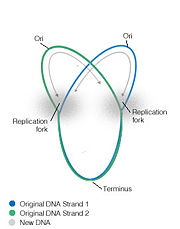
proteopedia linkproteopedia linkIn most bacterial DNA replication initiation occurs at an origin where, due to the circular nature of the chromosome, the replication forks move bidirectionally to end at approxiametly 180 degrees away, at a specific sequence termini region [1]. Bacterial replication termination systems have been well studied in Eschericia coli and Bascillus subtilis. In both systems a trans-acting replication termination protein binds to a specific cis-acting DNA sequences, the replication termini (ter), and the DNA-protein complex arrests the progression of replication forks. The terminator sites are orientated so that protein binding is asymmetric, allowing the complexes to block the replication machinery from only one direction while letting them proceed unimpeded from the other direction [2]. In this way they are said to act in a polar manner. The proteins involved in this termination are non-homologous and differ structurally in E.coli and B.subtilis, although each contains similar contrahelicase activity and performs similar functions in arresting replication.

Bacterial replication fork
Termination (ter) Sites
Replication is terminated in bacterial systems such as E.coli and B.subtilis by a "replication fork trap", studded with termination sites which causes the bidirectional forks to pause, encounter and fuse within a region called the terminus region. In E.coli the termination regions are spread across nearly half the chromosome compared to B.subtilis where they cover only ~10%. Termination regions are made up of two groups, opposite to each other, containing inverted sequences for the polar arrest of the replication helicase. In E.coli the 5 ter sites, J, G, F, B and C are arranged opposed to ter sites H, I, E, D and A, and can arrest the fork progressing in the clockwise direction and can block the anticlockwise direction, respectively. The replication fork progressing in a clockwise direction will encounter the terC site first and pause. If the fork progressing from the anticlockwise direction meets the clockwise fork while paused, replication is terminated, however if it does not meet its anti-fork it will proceed until it reaches the next termination site, terB, where it will pause again, etc [8]. Therefore multiple ter sites are important as infrequently utilized backups, to ensure that the fork does not leave the terminus region, and that termination is completed. Multiple regions to entrap the replication fork means that if an inactivating mutation arises within a ter site, then arrest can still occur at another ter sequence [6].
Replication Terminator Protein (Bacillus subtilis)
| Replication Termination Protein (RTP), found in Bacillus subtilis, is a member of the ‘winged helix’ protein family, and terminates bacterial DNA replication by arresting the replication forks through interactions with DNA in a sequence specific manner [9]. RTP blocks the replication fork through contrahelicase activity; the ability to specifically inhibit the helicase replication machinery and has an additional role in arresting transcription [1][2]. In B. subtilis the bipartite ter sequence is overlapping, and each inverted repeat contains core (IRIB) and an auxillary (IRIA) sites. RTP binds to these sequences, resulting in the impediment the replication fork helicase.
RTP Structure
Active RTP is a homodimer composed of 14.5 kDa subunits. The structure of the protein has been determined to a 2.6 A resolution using X-ray crystallography [3]. The RTP protein contains three major structural domains for its specific functionality; DNA-binding,
and dimer-dimer interaction domains [3]. The RTP is organized into a dimer by the association of their
within the C-terminus [3]. The ‘’ is believed to be involved as the major DNA-binding domain; with the two helices slotting into the major grove and two β strands inserting into the minor grove [14]. This binding interaction is vastly different from the Tus-ter interactions.
RTP Mechanism of Action
An RTP dimer binds the core sequence and the complex formed allows a second dimer to cooperatively to bind to the auxiliary site. In the absence of a core site, the auxiliary site is unable to bind RTP. Furthermore, without the auxiliary site, RTP is unable to block the replication fork, as the interaction of both dimers has been suggested to provide enough DNA binding strength to displace the replication fork. This binding explains how the symmetrical RTP can block replication helicase machinery in an asymmetric manner. The blocking end occurs at the core site, while it is believed that the non-blocking auxiliary site may let replication through as there is less contact points of the dimer to the DNA and the replication machinery coming from this direction is predicted to displace the dimer that is weakly bound to the auxiliary site, which would then displace the dimer bound to the core [10]. Biochemical and mutational studies have identified particular residues that are vital for the functionality of RTP. Mutations within a hydrophobic region at residues Glu-30 and Tyr-33 causes the loss of contrahelicase ability [10]. These mutations do not affect dimer-dimer interactions or DNA binding activity and indicate that simple DNA binding is not able to block the replication fork. This provided evidence that RTP and the replication fork machinery interact specifically [10].
|
The Terminus Utilization Substance (Escherichia coli )
The E.coli protein that is responsible for termination is a 36kDa protein named Tus (Terminius Utilization Substance) that binds 23bp ter sites and arrests the replication helicase, DnaB, responsible for separating the two strands of DNA []. Unlike RTP termination sites, the ten E.coli ter sites do not contain inverted sequences or direct repeats and Tus binds as a monomer to a highly conserved core region of 13bp [8]. The tus-ter complex is known to terminate replication by arresting the replication machinery in a in a polar manner however there is great discrepancy in evidence whether Tus specifically interacts or physically blocks the DnaB helicase to arrest its progression [1].
Tus structure
Tus is a member of the replication termination protein family and has no similarity to any DNA-binding motif that is known, and is organized into two discontinuous domains ( and
) that consist of α helical and β sheets. Two pairs of antiparallel β strands link the N and C terminal protein domains and form the . This provides a large, positivley charged, central cleft into which the double helix (deformed locally) can fit, so that the two α helical domains flank the DNA. The interdomain β strands, which makes up the core region of the 13bp DNA-binding, that sit across from the DNA, accesses a deepened major groove and contacts several of the bases within this groove, and is responsible for ter sequence recognition and binding to DNA. There are 17 sequence-specific interactions between the DNA and the α helixes and β strands presented by Tus, although the majority of these interactions are from the proximinal β sheets. The helicase-blocking or , of Tus consists of α helices and loops from N and C terminal domains. Tus is unrelated structurally to the replication termination protein despite their similar functions.
Tus Mechanism of Action
Two models were proposed to explain the mechanism of Tus activity generated by early experiments. The "clamp model" proposed that the Tus-ter complex created a barrier that arrested the progression of the replication machinery from one direction but not the other, by DNA binding [12]. The "interaction model" suggested that a particular region of Tus specifically interacted with the progressing helicase, causing it to halt the fork, and this interaction would only be possible at one face of the protein [13]. The structure of the tus-DNA complex has recently been solved [8]. It suggests that the protein can block helicase approaching from one direction and not the other, without the necessity of specific Tus–helicase interactions. The Tus-Ter complex could act as a physical barrier against the replication fork at the non-permissive face; the α helical regions protrude from the protein around the DNA and block the helicase from accessing the region tightly bound to the DNA. On the other hand, when the helicase advances from the opposite direction it does not encounter the α helical barriers and can disrupt Tus-DNA binding by interrupting the interdomain β strands, causing Tus to be released. This simple model is supported by studies where mutants were screened after exhibiting a reduction in their ability to arrest replication [8]. Most of the mutations occurred in the interdomain β-strands and none of these mutations occurred in the blocking surface that may contact the progressing helicase [8]. However it is important to note that a specific interaction between the blocking face and the helicase cannot be ruled out based on structural studies, and that it if present it may have a role to enhance the physical barrier’s effectiveness.
| |
Biological Significance
The role of the replication fork arrest was primarily believed to be of great importance for the faithful termination of replication, segregation of chromosomes and faithful inheritance of a stable genome. However recent studies where the rtp and tus genes of B.subtilis and E.coli, respectively, were knocked out, suggested that this role is dispensable. Indeed, bacterial systems that have mutations within these genes can survive in the environment and appear identical in both growth rate and cell morphology compared to wildtype bacteria, suggesting that replication termination is not a requirement for cytokinesis [4]. It has recently been suggested that this form of termination may have roles in aiding the co-ordination and optimization of recombination events preceding replication in bacteria, and preventing over-replication. It is also suggested that termination may occur by specific dif sites, conserved sites that are located near the terminus region that are involved in homologous recombination. In fact the dif-terminus hypothesis proposes that termination occurs at or near these sites, where after termination of the replication forks, the dif-sites would undergo site-specific recombination, and that this would resolve the dimer chromosomes and complete replication
References

