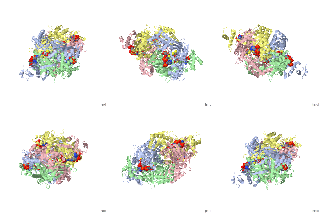
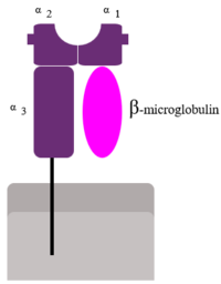
Beta two microglubulin in human class I major histocompatibility complex (MHCb2m)
Human β2-Microglobulin is the non-covalently bound
of the human class I
major histocompatibility complex (MHC-I). Its function is to ensure proper folding and cell-surface expression of MHC-1.
The natural turnover of MHC-I gives rise to the release of b2m into plasmatic fluids at ~0.1
um and to its catabolism in the kidney. In case of renal dysfunction, b2m concentration increases
up to 60-fold, giving rise to pathogenic accumulation of filamentous structures, displaying the
typical properties of amyloid fibrils, principally in the joints and connective tissue.
Monomeric human b2m (Mhb2m)
The first crystal structure of Monomeric human b2m (Mhb2m) is solved in 2002 (pdb 1LDS). The protein is 99 residue in length and has a seven-stranded β sandwich fold typical of the Immunoglobulin superfamily. It is stabilized by a single disulfide bond between
Cys-25 and Cys-80, which links the two β sheets.
Structural comparison of MHCb2m and Mhb2m
Both of the two strucures adopt seven-stranded β sandwich fold with a short C' β strand located in the loop connecting strands C
and D. The most significant difference in the ctrystal structures of Mhb2m and MHCb2m involves residues in β strand D and the succeeding loop. When complexed with the MHC heavy chain, residues 50-56 of MHCb2m form two short β strands that separated by a two residue β bulge. These strands (depicted as D1 and D2 in Fig.1) each forms three main-chain-main-chain hydrogen bonds to the adjacent β strand E. The bulge in MHCb2m effectively twists the edge strand, which facilitate its binding to the surface of the heavy chain. However, this β bulge no longer exits in the crytal strucure of Mhb2m. The in Mhb2m provides an ideal assembly surface, making this edge-strand pair vulnerable to aggregation. The hydrogen-bonding potential of strand D is satisfied by the formation intermolecular interactionswith adjecent molecules, demonstrating the potential for this region to propagate assembly through edge-strand interactions.
In addition, the changes observed in strand D result in differenr orientations of the side chains of residues 50-54. As a result, His-51 (which points inwards in the structure of MHCb2m) rotates by approximately 180, such that it now points away from the hydrophobic core of the protein. This would remove the second protevtive feature from the edge strand, facilitating further interaction in this region (Fig.2).
Amyloid Fibril formation of Mhb2m
Mhb2m fiber formation conditions
While the exact factors that cause b2m fibril formation in vivo are not known, several means exist to generate b2m amyloid fibrils in vitro. b2m amyloid fibrils can be generated under acidic conditions (pH < 3.6), by truncating the first six N-terminal amino
acids, by dialyzing the protein into distilled water followed by membrane drying, by mixing the protein with collagen at pH=6.4, by sonicating the protein in the presence of sodium dodecyl sulfate at pH=7.0, and by incubating the full-length protein at
physiological conditions in the presence of stoichiometric amounts of Cu(II).The latter method is particularly intriguing and may have relevance to the in vivo process because of the very near physiological conditions used.
Mhb2m fiber formation mechanism at neutral pH
A structural trigger
Alterations of the protein sequence have been used to stimulate the formation of fibrils at neutral pH. Specifically, truncation of six residues from the N-terminal region(DN6), as well as mutation in this region (P5G) or in the B/C or D/E loops (
P32A, P32G, D59P) of the protein all enhance its ability to form amyloid in vitro, while substitutions elsewhere in the protein have little effect. These studies have the common feature that they encourage partial unfolding of b2m, allowing the aggregation-prone regions of the polypeptide sequence to be exposed and to participate in intermolecular interactions.
The b2m variants, P32G and P32V, have been used to show that wild-type b2m populates a native-like folding intermediate (called
IT) en route to the native state that contains a non-native trans Pro32 bond. Increased population of this intermediate in the variant P32G was shown to be concomitant with increased ability of the protein to elongate b2m fibril seeds. Other variants, such as P5G and DN6, also affect isomerisation of the Pro32 peptide bond, promoting population of IT and enabling fibril nucleation at pH 7.0.
In separate studies b2m has been shown to form oligomers and fibrils at neutral pH by addition of Cu2+ and 1 M urea. This induces the formation of a non-native species, called M*, that appears identical to IT by size exclusion chromatography (SEC). Coordination of the metal ion promotes peptide bond isomerisation at Pro32, and the subsequent rapid formation of oligomers as
judged by SEC, providing further supporting evidence that the isomerisation of Pro32 is a key initial step on the pathway to fibrils.Structural studies of the trapped folding intermediate, IT (orM*), have provided molecular insights into the possible aggregation mechanisms of b2m at neutral pH. For example, crystallographic data for the mutant, P32A shows that this conformer results in substantial reorganisation of aromatic side-chains that are present in the core of the native protein. In this structure, a number of aromatic and hydrophobic residues are displaced including Phe30, Phe56, Trp60, Phe62, Tyr63 and Leu54. These rearrangements present a strip of exposed hydrophobic residues on the protein surface, providing a possible avenue for protein aggregation (Fig.3). This species crystallises as a dimer underlining its potential to form intermolecular interactions.
Fibrillar architecture
In aqueous solution at neutral or acidic condition, amyloid-like fibrils are formed from b2m that show a long-straight, left-hand twisted and unbranched morphology when observed by EM and AFM (Fig.4)
A unifying mechanism of b2m fibril formation
One of the most striking observations of the b2m assembly pathways is that the fibrils formed commencing from a highly
denatured state or a native-like precursor are apparently indistinguishable, suggesting that their assembly pathways must converge
to a similar fibrillar product. This could occur by unfolding of native b2m to allow reorganisation of the protein structure; or by
refolding of the highly dynamic polypeptide chain at pH 2.5 to a more structurally ordered intermediate species (Fig.5).
Similarities between the assembly mechanisms are supported by several reasons:
- . The proposal that aromatic interactions are crucial for driving fibrillogenesis under both sets of conditions.
- . Another convergent feature arises from a suggested model for the fibrils formed at neutral pH that contains a highly charged surface, which could be neutralised at low pH, perhaps allowing for a convergence of the mechanisms of assembly at acidic and neutral pH and explaining why the kinetics of fibril formation are much more rapid under acidic conditions.
- .An additional common characteristic is that many variants capable of fibril formation at neutral pH have the effect of destabilising the Nterminal region of the protein, which is also highly unfolded in the structural ensembles formed at low pH.
- . The role of trans Pro32 in fibril formation at low pH is currently unknown, although 80% of the molecules would be expected to contain the trans conformation in the acid denatured state. Moreover, the observation that the rate of fibril formation of P32G is similar to that of wild-type b2m when studied at pH 2.5 is suggestive of a common trans amyloid precursor.


