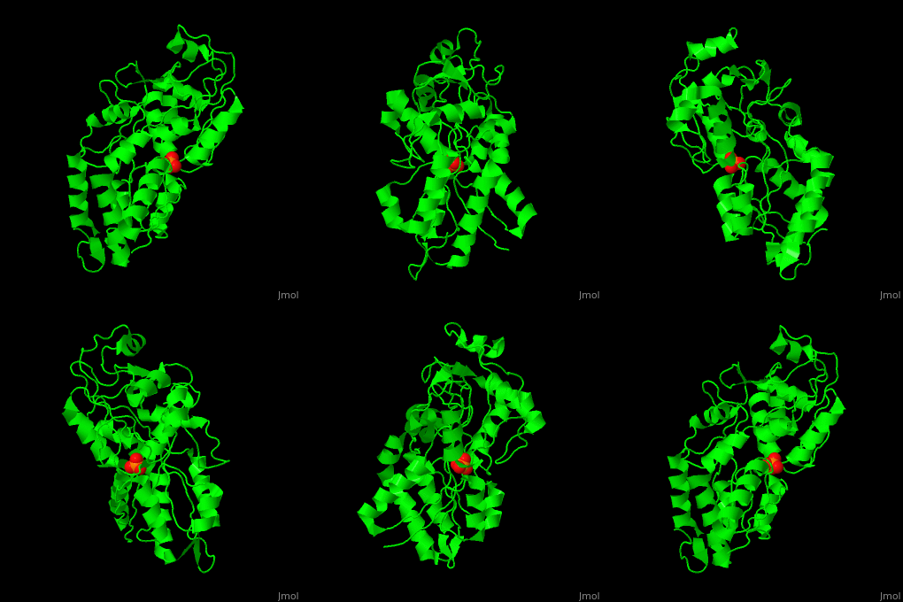
Phosphate-binding protein
From Proteopedia
| |||||||||||
3D structures of phosphate-binding protein
Updated on 22-September-2020
2v3q – hPBP + Pi – human
1qui, 1oib - EcPBP (mutant) - Escherichia coli
1pbp, 2abh, 1ixh – EcPBP + Pi
1qui - EcPBP (mutant) + Br + Pi
1quj, 1qul - EcPBP (mutant) + Cl + Pi
1quk, 1ixi, 1ixg, 1a40 - EcPBP (mutant) + Pi
1a54, 1a55 - EcPBP (mutant) + dihydrogenphosphate
1pc3, 4lvq – PBP + Pi – Mycobacterium tuberculosis
3w9v, 3w9w – upPBP + Pi – unidentified prokaryote
4m1v - upPBP (mutant) + Pi
4exl, 4h1x – SpPBP – Streptococcus pneumoniae
4lat – SpPBP + Pi
4omb, 4pqj – PBP + Pi – Pseudomonas aeruginosa
5wnn – PBP + Pi – Brucella melitensis
5ub3 – XcPBP – Xanthomonas citri
5i84, 5ub4 – XcPBP + Pi
5ub6 – XcPBP + PPi
5ub7 – XcPBP + ATP
5j1d – PBP + Pi – Stenotrophomonas maltophilia
4jwo – PBP – Planctomyces limnophilus
References
- ↑ Gonzalez D, Richez M, Bergonzi C, Chabriere E, Elias M. Crystal structure of the phosphate-binding protein (PBP-1) of an ABC-type phosphate transporter from Clostridium perfringens. Sci Rep. 2014 Oct 16;4:6636. doi: 10.1038/srep06636. PMID:25338617 doi:http://dx.doi.org/10.1038/srep06636
- ↑ Wang Z, Choudhary A, Ledvina PS, Quiocho FA. Fine tuning the specificity of the periplasmic phosphate transport receptor. Site-directed mutagenesis, ligand binding, and crystallographic studies. J Biol Chem. 1994 Oct 7;269(40):25091-4. PMID:7929197

