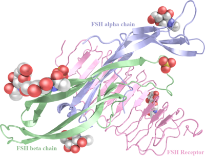
Human Follicle-Stimulating Hormone Complexed with its Receptor
From Proteopedia
| For the date when the most recent work on this article was done, click on the history tab above. |
NEEDS UPDATING TO INCLUDE 4ay9.
Contents |
Crystal Structure of Human Follicle-Stimulating Hormone Complexed with its Receptor
Follicle-stimulating hormone (FSH) is central to reproduction in mammals. It acts through a G-protein-coupled receptor on the surface of target cells to stimulate testicular and ovarian functions. We present here the 2.9-A-resolution structure of a partially deglycosylated complex of human FSH bound to the extracellular hormone-binding domain of its receptor (FSHR(HB)). The hormone is bound in a hand-clasp fashion to an elongated, curved receptor. The buried interface of the complex is large (2,600 A2) and has a high charge density. Our analysis suggests that all glycoprotein hormones bind to their receptors in this mode and that binding specificity is mediated by key interaction sites involving both the common alpha- and hormone-specific beta-subunits. On binding, FSH undergoes a concerted conformational change that affects protruding loops implicated in receptor activation. The FSH-FSHR(HB) complexes form dimers in the crystal and at high concentrations in solution. Such dimers may participate in transmembrane signal transduction.
Structure of human follicle-stimulating hormone in complex with its receptor., Fan QR, Hendrickson WA, Nature. 2005 Jan 20;433(7023):269-77. PMID:15662415
From MEDLINE®/PubMed®, a database of the U.S. National Library of Medicine.
Disease
| ||||||||||
| ||||||||||
Known disease associated with this structure: Follicle-stimulating hormone deficiency, isolated OMIM:[136530], Ovarian dysgenesis 1 OMIM:[136435], Ovarian hyperstimulation syndrome OMIM:[136435], Ovarian response to FSH stimulation OMIM:[136435], Ovarian sex cord tumors OMIM:[136435]
About this Structure
1XWD is a 6 chains structure of sequences from Homo sapiens. Full crystallographic information is available from OCA.
The leucine-rich domain of the receptor ; leucine-rich repeats often are horseshoe- or arc-shaped.
Add about cysteine-knot.
Add about seat belt.
Keep in mind this is just the leucine-rich repeat, amino-terminal portion of the receptor and that in the full receptor structure part of the chain also forms a G protein-coupled receptor and a brief intracellular domain at the C-terminus.
References
- Fan QR, Hendrickson WA. Structure of human follicle-stimulating hormone in complex with its receptor. Nature. 2005 Jan 20;433(7023):269-77. PMID:15662415 doi:10.1038/nature03206
- Jiang X, Liu H, Chen X, Chen PH, Fischer D, Sriraman V, Yu HN, Arkinstall S, He X. Structure of follicle-stimulating hormone in complex with the entire ectodomain of its receptor. Proc Natl Acad Sci U S A. 2012 Jul 31;109(31):12491-6. Epub 2012 Jul 16. PMID:22802634 doi:10.1073/pnas.1206643109
See also
- Leucine-rich repeat proteins
- Hormone
- Membrane proteins
- Receptor
- Transmembrane (cell surface) receptors
- G protein-coupled receptors
- 1hcn - human chorionic gonadotropin placental hormone
- 1hrp - human chorionic gonadotropin placental hormone
- 1hd4 - human chorionic gonadotropin placental hormone alpha subunit
- 3g04 - Human thyroid-stimulating hormone receptor in complex with a thyroid-stimulating autoantibody
3D structures of follicle-stimulating hormone
See Follicle-stimulating hormone
External Resources
- Sequence-Structure-Function-Analysis of Glycoprotein Hormone Receptors
- GRIS: Glycoprotein-hormone Receptors Information System
Page originally seeded at 1xwd by OCA on Tue Feb 17 21:34:49 2009

