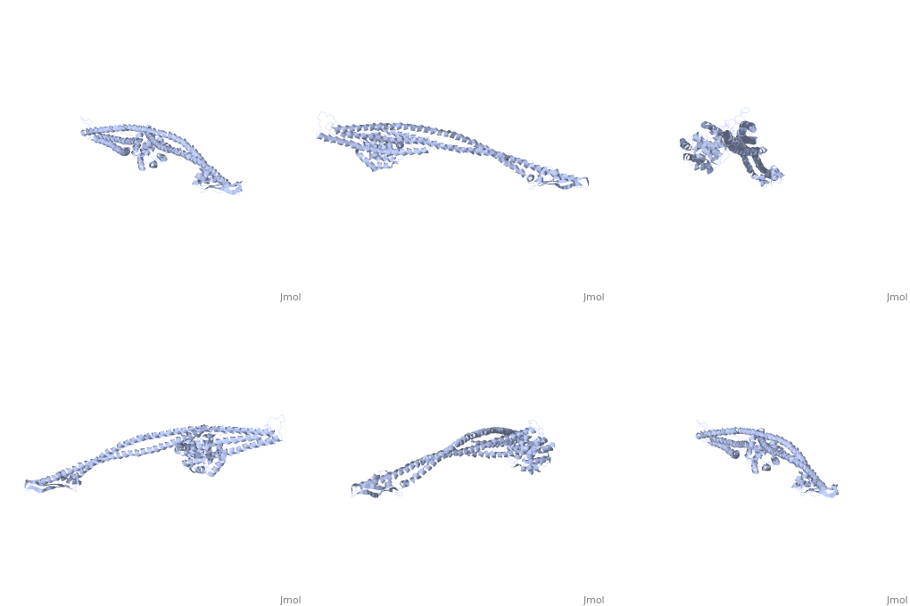
Colicin Ia is a type of Colicin, a bacteriocin made by E. coli which acts against other nearby E. coli to kill them by forming a voltage-dependent channel in the inner membrane of the cells that it targets.
Synthesis and release
Colicin Ia is a 69kDa protein encoded on a high molecular weight plasmid[1] that is induced by the SOS system during stress[2]. The gene is tightly linked to its specific Colicin Immunity Protein, Iia, to protect the colicinogenic cell from the cytotoxic activity of the colicin[3]. The gene goes not encode a lysis protein, like Colicin V for its release from the cell, instead it uses export proteins already present to push itself out of the cell and not destroy it.
Colicin Ia has a high degree of homology to Colicin Ib, and is antigenically similar to it as well[4].
Mechanism of uptake
(PDB entry 2hdi).
The flexible[5] N terminus of ColIa is responsible for binding to the colicin I receptor (Cir), with high affinity (Kassoc=1x10¹⁰M-1[6]). Cir is a 22-stranded plugged β barrel TonB-dependent transporter. Its normal function on E. coli is binding and transporting Fe3+ across the outer membrane, but is parasitized by colicins for their transport and entry. When ColIa binds to Cir it results in a big conformational change, resulting in Cir being open and exposed extracellularly, with the ColIa R-domain positioned directly above it, bound at approximately 45o to the membrane. This results in the T and C domains remaining far above the membrane and away from the receptor. If Cir is indeed the molecule that transports ColIa across the membrane, this conformational change is that that would be required for penetration by the colicin[7]. ColIa molecules with the receptor binding domain deleted are still able to enter cells, although less effectively, as Cir weakly binds to the translocation domain as well as the receptor binding domain[8].
The entry mechanism for ColIa into the periplasm after receptor binding is still unclear. The pair of α helices that separate the T and C domains from the R domain was suggested to allow ColIa to span the membrane, with the R domain being left outside the cell. The C domain is the only one expected to pass across the inner membrane and enter the cytoplasm[9]. Alternatively the 45o angle made with the membrane may allow the ColIa to search the surrounding regions for a co-transporter or other method of entry to the cell. For killing by ColIa to be successful, Cir requires a functional TonB box sequence, as does ColIa, which implies that an interaction with TonB is required for uptake of ColIa[10].
ColIa has been shown to transport cargo proteins on its N terminus across the lipid bilayer when it penetrates the target cell. This transport uses the voltage across the bilayer to bring the folded proteins across the membrane[11].
Some strains of E. coli are resistant to killing by ColIa, such as those lacking the Cir receptor protein. Colicin Ia is then susceptible to fragmentation by outer membrane proteins of the cell that it is targeting, a process not mediated by Cir[12].
Killing Activities
The 177 residue Pore Formation domain, once it has translocated across the periplasmic space, inserts into the inner membrane of the target E. coli and forms a voltage-dependent ion channel that opens at a positive potential and closes at a negative potential, and conduct cations and anions[13]. ColIa transfers a region of itself containing 4 α helices (H6, H7, H8 and H9) across the membrane as part of its gating[14]. Parts of H8 and H9 form the lining of the channel, with the residues with the greatest effects of conductance and selectivity being placed near the cis end of the channel[15].
The structural and mechanical sides of the pore formation by ColIa is an enigma; structurally there isn't enough protein to form such a pore - each helix is approximately 12 residues long[16], and mechanically the gating involves the transmembrane movement of a large portion of the protein, but it is unclear how this senses the voltage or forms the pore.
Insertion of ColIa into the lipid membrane triggers a series of conformational change across the whole of ColIa to form the ion channel[17], probably through a series of intermediates. While a monomer of ColIa in the membrane is sufficient to form the channel and kill the cell, studies have shown that the channel, when enough molecules are present, exists as a multimer with a larger pore diameter[18][19].
Once the pore has inserted into the membrane and is functioning fully, it inhibits the active transport of a number of molecules, primarily proline, thiomethyl-β-D-galactoside, and K+ ions. Not only does it prevent their uptake, it can also trigger the rapid release of the molecules. It also inhibits succinate uptake, which accounts for the inhibition of respiration observed in targeted cells, and has an effect on the uptake of glycerol[20]. All of this leads to an uncoupling of electron transport from active transport, and affects energy metabolism with its interference with the membrane, leading to a depolarisation of the membrane[21][22][23].

