Fadel A. Samatey Group
From Proteopedia
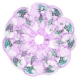 | |
| Crystal structure of flagellar hook fitted into electron density map obtained by cryo-electron microscopy[1]. |
Fadel A. Samatey was Head of the Transmembrane Trafficking Unit at the Okinawa Institute of Science and Technology (OIST) (Japan), 2007-2016. Samatey's group uses X-ray crystallography, cryo-Electron microscopy, genetic, and biochemical approaches to elucidate the structures and functions of proteins, especially type III secretion proteins in bacterial flagella.
From 1996-2007 Samatey was a member of the Keiichi Namba Group, from 1996 at Matsushita Electric, from 1997 in the ERATO Protonic Nanomachine Project, and from 2002 at Osaka University, Japan. Samatey earned his Ph.D. in 1992 at Université Joseph Fourier in Grenoble, France.
Below are listed contributions from the Samatey Group, most recent first.
Contents |
Interests and Objectives
Motility is a very important function in the living world. For this purpose, organisms such as bacteria have developed the most incredible molecular machine: the flagellar system. Bacteria such as Escherichia coli and Salmonella typhimurium swim by rotating long helical filaments called the flagellum. The flagellum is a complex structure made by the association of many different proteins. It can be divided into three parts: 1) the filament: a long, rigid, tubular structure that works as a helical propeller, 2) the hook: a short, highly flexible tubular segment that works as a universal joint, and 3) the basal body: a rotary motor embedded in the cell membrane.
During the assembly of the flagellum, all the flagellar axial proteins are exported from the cytoplasm to the flagellum distal end through a 2 -3 nm channel located at its centre. This export mechanism is regulated by a specialized protein export system located on the cytoplasmic side of the basal body. It is called the Type III Export Apparatus and is found throughout the bacterial kingdom. In the case of Salmonella, this export apparatus is made by six membrane proteins: FlhA, FlhB, FliO, FliP, FliQ, FliR, and three cytoplasmic proteins: FliI, FliH and FliJ. The export apparatus of the bacterial flagellum is homolog to the type III secretion system (T3SS) found in Gram-negative pathogenic bacteria. The T3SS role is to secrete virulence factors to host cells, leading to diverse diseases. To understand both the bacterial flagellum and its export apparatus, we have been doing structural studies on some flagellar proteins and genetic studies on the export apparatus.
Contact: fadel.samatey at gmail.com
Contributions from 2011 to Present
- Bulieris PV, Shaikh NH, Freddolino PL, Samatey FA. Structure of FlgK reveals the divergence of the bacterial Hook-Filament Junction of Campylobacter. Sci Rep. 2017 Nov 16;7(1):15743. doi: 10.1038/s41598-017-15837-0. PMID:29147015 doi:http://dx.doi.org/10.1038/s41598-017-15837-0
- Ly T, Krieger I, Tolkatchev D, Krone C, Moural T, Samatey FA, Kang C, Kostyukova AS. Structural Destabilization of Tropomyosin Induced by the Cardiomyopathy-linked Mutation R21H. Protein Sci. 2017 Nov 6. doi: 10.1002/pro.3341. PMID:29105867 doi:http://dx.doi.org/10.1002/pro.3341
- Barker CS, Meshcheryakova IV, Kostyukova AS, Freddolino PL, Samatey FA. An intrinsically disordered linker controlling the formation and the stability of the bacterial flagellar hook. BMC Biol. 2017 Oct 27;15(1):97. doi: 10.1186/s12915-017-0438-7. PMID:29078764 doi:http://dx.doi.org/10.1186/s12915-017-0438-7
The “ID-Rod-Stretch” is an intrinsically disordered linker found in both FlgE and FlgG, the proteins that respectively make the hook and the distal rod of the bacterial flagellum. Experiments done in FlgE of Salmonella enterica and of Campylobacter jejuni reveal the role of the ID-Rod-Stretch in the formation and stability of the flagellar hook. See the special video abstract. See results in interactive 3D.
- Matsunami H, Barker CS, Yoon YH, Wolf M, Samatey FA. Complete structure of the bacterial flagellar hook reveals extensive set of stabilizing interactions. Nat Commun. 2016 Nov 4;7:13425. doi: 10.1038/ncomms13425. PMID:27811912 doi:http://dx.doi.org/10.1038/ncomms13425
The bacterial flagellar hook, which is made by the polymerization of multiple copies of FlgE protein, is a flexible segment connecting the flagellar filament to the motor. The structure presented in this article puts in evidence the complex web of interactions between FlgE molecules. These interactions stabilize the flagellar hook during its function as a universal joint. See results in interactive 3D.
- Yoon YH, Barker CS, Bulieris PV, Matsunami H, Samatey FA. Structural insights into bacterial flagellar hooks similarities and specificities. Sci Rep. 2016 Oct 19;6:35552. doi: 10.1038/srep35552. PMID:27759043 doi:http://dx.doi.org/10.1038/srep35552
The assembly of about a hundred molecules of FlgE protein makes the bacterial flagellar hook. FlgE has a high variability in amino acid residues composition and in molecular weight. Our study shows that FlgE can be divided in two distinct parts. The first part comprises domains that are found in all FlgE proteins and will make the basic structure of the hook common to all flagellated bacteria. The second part, hyper-variable both in size and structure, will be bacteria dependent and will give to the hook additional and specific properties to the bacterium its belong to. See results in interactive 3D.
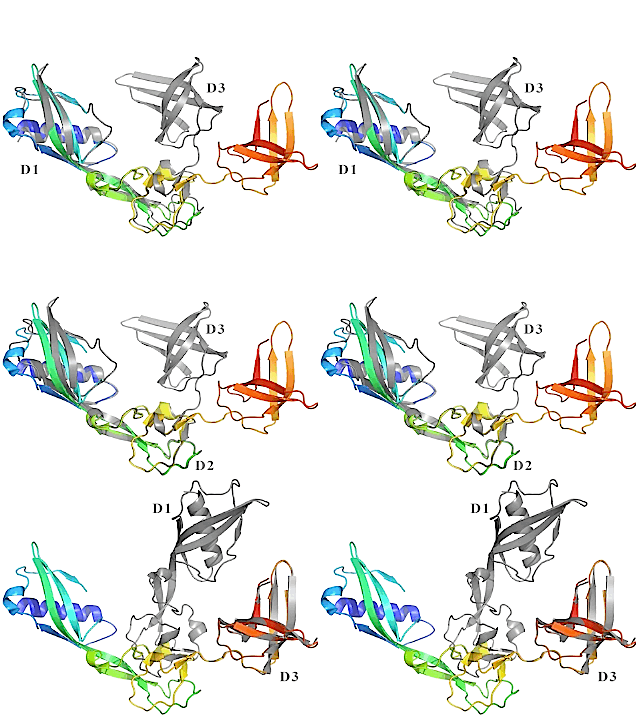 | |
| Superposition of the different domains of the open and closed forms of FlgA. See it in interactive 3D. |
- Matsunami H, Yoon YH, Meshcheryakov VA, Namba K, Samatey FA. Structural flexibility of the periplasmic protein, FlgA, regulates flagellar P-ring assembly in Salmonella enterica. Sci Rep. 2016 Jun 7;6:27399. doi: 10.1038/srep27399. PMID:27273476 doi:http://dx.doi.org/10.1038/srep27399
The bacterial flagellar P-ring is located in the periplasm and is important for the externalization of the bacterial flagellum in Gram-negative bacteria. FlgA is a protein that regulates the assembly of the P-ring. Deletion of flgA gene leads to non-flagellated cells. This study showed that it is possible, by reducing the flexibility of FlgA, to block the formation of the flagellum. See results in interactive 3D.
- Barker CS, Inoue T, Meshcheryakova IV, Kitanobo S, Samatey FA. Function of the conserved FHIPEP domain of the flagellar type III export apparatus, protein FlhA. Mol Microbiol. 2016 Apr;100(2):278-88. doi: 10.1111/mmi.13315. Epub 2016 Feb 10. PMID:26691662 doi:http://dx.doi.org/10.1111/mmi.13315
FlhA is the largest membrane protein of the Type III export apparatus and is homologous to the large family of FHIPEP export proteins. FHIPEP proteins contain a highly-conserved cytoplasmic domain. This study suggested that the FHIPEP region of FlhA is part of the gate regulating substrate entry into the export apparatus pore.
- Barker CS, Meshcheryakova IV, Inoue T, Samatey FA. Assembling flagella in Salmonella mutant strains producing a type III export apparatus without FliO. J Bacteriol. 2014 Dec;196(23):4001-11. doi: 10.1128/JB.02184-14. Epub 2014 Sep 8. PMID:25201947 doi:http://dx.doi.org/10.1128/JB.02184-14
Deletion of the fliO gene creates a mutant strain that is poorly motile; however, suppressor mutations in the fliP gene can partially rescue motility. Here we found a missense mutation that localized to the clpP gene, which encodes the ClpP subunit of the ClpXP protease, and a synonymous mutation that localized to the fliA gene, which encodes the flagellar sigma factor, σ(28) also could partially rescue motility of fliO mutant strains. Combining these suppressor mutations with mutations in the fliP gene fully rescued flagellar biosynthesis and motility for fliO deletion mutant strains. The suppressor mutations in the fliP gene had the greatest effect. This suggests that the function of FliO is closely associated with regulation of FliP during assembly of the flagellum.
- Mise T, Matsunami H, Samatey FA, Maruyama IN. Crystallization and preliminary X-ray diffraction analysis of the periplasmic domain of the Escherichia coli aspartate receptor Tar and its complex with aspartate. Acta Crystallogr F Struct Biol Commun. 2014 Sep;70(Pt 9):1219-23. doi:, 10.1107/S2053230X14014733. Epub 2014 Aug 27. PMID:25195895 doi:http://dx.doi.org/10.1107/S2053230X14014733
- Barker CS, Meshcheryakova IV, Sasaki T, Roy MC, Sinha PK, Yagi T, Samatey FA. Randomly selected suppressor mutations in genes for NADH:quinone oxidoreductase-1, which rescue motility of a Salmonella ubiquinone-biosynthesis mutant strain. Microbiology. 2014 Apr 1. doi: 10.1099/mic.0.075945-0. PMID:24692644 doi:http://dx.doi.org/10.1099/mic.0.075945-0
This study showed amino acid substitutions at or near to the surface of NADH:quinone oxidoreductase-1 (NDH-1) increased growth, motility and flagellar biogenesis of mutant strains unable to synthesize the aerobic respiratory chain electron carrier ubiquinone. Therefore, NDH-1 activity in Salmonella is important for flagellar biogenesis.
- Galeva A, Moroz N, Yoon YH, Hughes KT, Samatey FA, Kostyukova AS. Bacterial flagellin-specific chaperone FliS interacts with anti-sigma factor FlgM. J Bacteriol. 2014 Mar;196(6):1215-21. doi: 10.1128/JB.01278-13. Epub 2014 Jan 10. PMID:24415724 doi:http://dx.doi.org/10.1128/JB.01278-13
- Stadler AM, Unruh T, Namba K, Samatey F, Zaccai G. Correlation between supercoiling and conformational motions of the bacterial flagellar filament. Biophys J. 2013 Nov 5;105(9):2157-65. doi: 10.1016/j.bpj.2013.09.039. PMID:24209861 doi:http://dx.doi.org/10.1016/j.bpj.2013.09.039
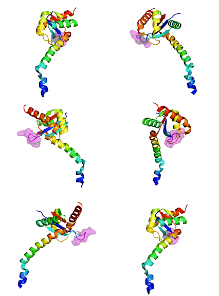 | |
| FlhBc of Salmonella (3b0z). Small surface is loop 281-285. See it in interactive 3D. |
- Meshcheryakov VA, Barker CS, Kostyukova AS, Samatey FA. Function of FlhB, a membrane protein implicated in the bacterial flagellar type III secretion system. PLoS One. 2013 Jul 11;8(7):e68384. doi: 10.1371/journal.pone.0068384. Print 2013. PMID:23874605 doi:http://dx.doi.org/10.1371/journal.pone.0068384
Replacing the FlhB gene (see below) in Salmonella with that of Aquifex reduced motility. Mutations that restore motility appear to increase conformational flexibility of FlhB, providing further support for the hypothesis that such flexibility is crucial to its function.
- Meshcheryakov VA, Kitao A, Matsunami H, Samatey FA. Inhibition of a type III secretion system by the deletion of a short loop in one of its membrane proteins. Acta Crystallogr D Biol Crystallogr. 2013 May;69(Pt 5):812-20. doi:, 10.1107/S0907444913002102. Epub 2013 Apr 11. PMID:23633590 doi:10.1107/S0907444913002102
Reports the atomic structure of the cytoplasmic domain of FlhB, a highly-conserved part of the secretion apparatus crucial to the assembly of bacterial flagella. The Salmonella typhimurium and Aquifex aeolicus structures are similar, while sequence identity is 32%. Deletion of a surface loop (281-285) that is not conserved inhibits function. This loop appears to promote crucial flexibility. See results in interactive 3D.
- Barker CS, Samatey FA. Cross-complementation study of the flagellar type III export apparatus membrane protein FlhB. PLoS One. 2012;7(8):e44030. doi: 10.1371/journal.pone.0044030. Epub 2012 Aug 29. PMID:22952860 doi:http://dx.doi.org/10.1371/journal.pone.0044030
Salmonella that synthesized a recombinant FlhB protein, made of the transmembrane region of Aquifex FlhB fused to the cytoplasmic domain of Salmonella FlhB, produced very few flagella. Suppressor mutations reducing the synthesis of ubiquinone for the respiratory chain or in the flhA gene increased flagella numbers, demonstrating a genetic interaction between FlhB and respiratory chain activity, and FlhB and FlhA.
- Matsunami H, Samatey FA, Nagashima S, Imada K, Namba K. Crystallization and preliminary X-ray analysis of FlgA, a periplasmic protein essential for flagellar P-ring assembly. Acta Crystallogr Sect F Struct Biol Cryst Commun. 2012 Mar 1;68(Pt 3):310-3. Epub, 2012 Feb 23. PMID:22442230 doi:10.1107/S1744309112001327
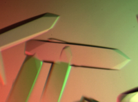 | |
| Crystals of the flagellar hook protein FlgE from C. jejuni produced in the Samatey lab[2]. |
- Kido Y, Yoon YH, Samatey FA. Crystallization of a 79 kDa fragment of the hook protein FlgE from Campylobacter jejuni. Acta Crystallogr Sect F Struct Biol Cryst Commun. 2011 Dec 1;67(Pt, 12):1653-7. Epub 2011 Nov 30. PMID:22139190 doi:10.1107/S1744309111043272
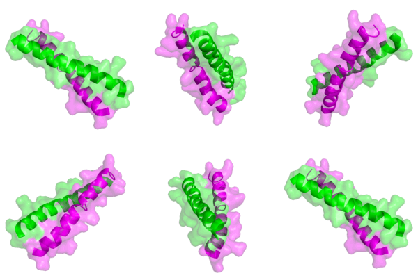 | |
| Coiled-coil of N-termini of chimeric non-muscle tropomyosin (3azd). |
- Meshcheryakov VA, Krieger I, Kostyukova AS, Samatey FA. Structure of a tropomyosin N-terminal fragment at 0.98 A resolution. Acta Crystallogr D Biol Crystallogr. 2011 Sep;67(Pt 9):822-5. Epub 2011, Aug 9. PMID:21904035 doi:10.1107/S090744491102645X
A high resolution atomic model of a chimeric (rat + yeast) N-terminal fragment of non-muscle tropomyosin confirms prior NMR results, but gives more accurate side-chain positions in the coiled-coil dimer.
- Meshcheryakov VA, Samatey FA. Purification, crystallization and preliminary X-ray crystallographic analysis of the C-terminal cytoplasmic domain of FlhB from Salmonella typhimurium. Acta Crystallogr Sect F Struct Biol Cryst Commun. 2011 Jul 1;67(Pt, 7):808-11. Epub 2011 Jun 30. PMID:21795800 doi:10.1107/S1744309111018938
- Meshcheryakov VA, Yoon YH, Samatey FA. Purification, crystallization and preliminary X-ray crystallographic analysis of the C-terminal cytoplasmic domain of FlhB from Aquifex aeolicus. Acta Crystallogr Sect F Struct Biol Cryst Commun. 2011 Feb 1;67(Pt, 2):280-2. Epub 2011 Jan 27. PMID:21301106 doi:10.1107/S1744309110052942
Contributions from 2001 to 2010
- Barker CS, Meshcheryakova IV, Kostyukova AS, Samatey FA. FliO regulation of FliP in the formation of the Salmonella enterica flagellum. PLoS Genet. 2010 Sep 30;6(9). pii: e1001143. PMID:20941389 doi:10.1371/journal.pgen.1001143
The membrane topology of FliO was determined and it has a small cytoplasmic domain whose overexpression partially bypassed the function of full-length FliO. Also, a fliO deletion mutant strain was almost non-motile, but motility was somewhat rescued by suppressor mutations in the fliP gene suggesting that FliO and FliP interact.
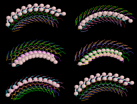 | |
| Simplified model of the rotating bacterial flagellar hook. |
- Furuta T, Samatey FA, Matsunami H, Imada K, Namba K, Kitao A. Gap compression/extension mechanism of bacterial flagellar hook as the molecular universal joint. J Struct Biol. 2007 Mar;157(3):481-90. Epub 2006 Oct 20. PMID:17142059 doi:10.1016/j.jsb.2006.10.006
Analyses the mechanism by which monomers of the flagellar hook slide against each other during hook rotation, to permit bending while transmitting torque.
- Kitao A, Yonekura K, Maki-Yonekura S, Samatey FA, Imada K, Namba K, Go N. Switch interactions control energy frustration and multiple flagellar filament structures. Proc Natl Acad Sci U S A. 2006 Mar 28;103(13):4894-9. Epub 2006 Mar 20. PMID:16549789 doi:10.1073/pnas.0510285103
Describes a massive molecular dynamics simulation that successfully accounts for polymorphic supercoiling in the bacterial flagellar filament. Interactions between protein monomer chains are dissected into permanent, sliding, and switch categories.
- Shaikh TR, Thomas DR, Chen JZ, Samatey FA, Matsunami H, Imada K, Namba K, Derosier DJ. A partial atomic structure for the flagellar hook of Salmonella typhimurium. Proc Natl Acad Sci U S A. 2005 Jan 25;102(4):1023-8. Epub 2005 Jan 18. PMID:15657146
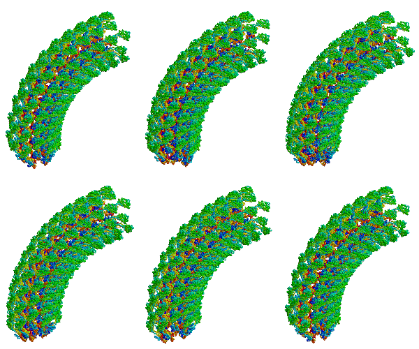 | |
| Bacterial flagellar hook, a molecular universal joint. |
- Samatey FA, Matsunami H, Imada K, Nagashima S, Shaikh TR, Thomas DR, Chen JZ, Derosier DJ, Kitao A, Namba K. Structure of the bacterial flagellar hook and implication for the molecular universal joint mechanism. Nature. 2004 Oct 28;431(7012):1062-8. PMID:15510139 doi:http://dx.doi.org/10.1038/nature02997
Reports the first structure of a major fragment of the protein monomer that assembles into the bacterial flagellar hook (1wlg, 1.8 Å resolution). Fits the monomer into an electron cryomicroscopic density map, resulting in straight and curved models of the hook, including a rotating model.
- Samatey FA, Matsunami H, Imada K, Nagashima S, Namba K. Crystallization of a core fragment of the flagellar hook protein FlgE. Acta Crystallogr D Biol Crystallogr. 2004 Nov;60(Pt 11):2078-80. Epub 2004, Oct 20. PMID:15502333 doi:10.1107/S0907444904022735
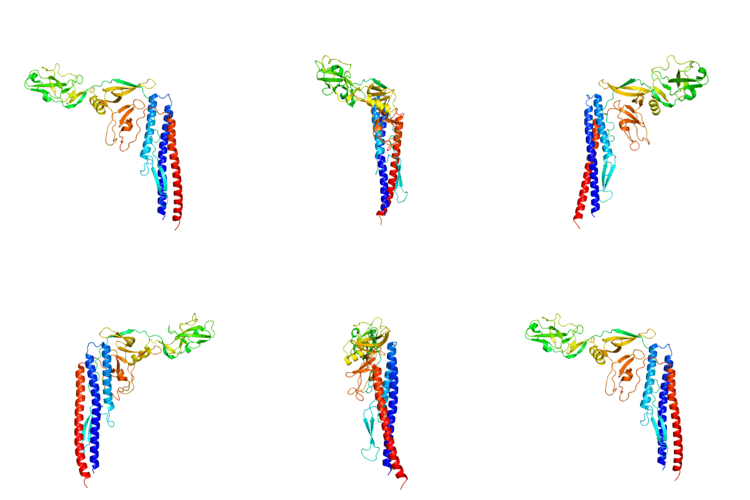 | |
| Monomer of the Salmonella flagellar filament, 1io1. |
- Samatey FA, Imada K, Nagashima S, Vonderviszt F, Kumasaka T, Yamamoto M, Namba K. Structure of the bacterial flagellar protofilament and implications for a switch for supercoiling. Nature. 2001 Mar 15;410(6826):331-7. PMID:11268201 doi:10.1038/35066504
Reports, for the first time, the atomic structure of a major fragment of the protein chain monomer that assembles into the bacterial flagellar filament (1io1, 2.0 Å resolution). The crystal contained chains of monomers, which revealed how the monomer protein chains fit together into protofilaments. Theoretical simulation revealed a possible mechanism for how the filament reverses direction, a mechanism crucial to how bacteria swim towards food or away from harm by reversing the flagellar motor.
Contributions from 1990 to 2000
- Samatey FA, Imada K, Vonderviszt F, Shirakihara Y, Namba K. Crystallization of the F41 fragment of flagellin and data collection from extremely thin crystals. J Struct Biol. 2000 Nov;132(2):106-11. PMID:11162732 doi:10.1006/jsbi.2000.4312
- Samatey FA, Xu C, Popot JL. On the distribution of amino acid residues in transmembrane alpha-helix bundles. Proc Natl Acad Sci U S A. 1995 May 9;92(10):4577-81. PMID:7753846
- Claros MG, Perea J, Shu Y, Samatey FA, Popot JL, Jacq C. Limitations to in vivo import of hydrophobic proteins into yeast mitochondria. The case of a cytoplasmically synthesized apocytochrome b. Eur J Biochem. 1995 Mar 15;228(3):762-71. PMID:7737175
- Lazzaroni JC, Vianney A, Popot JL, Benedetti H, Samatey F, Lazdunski C, Portalier R, Geli V. Transmembrane alpha-helix interactions are required for the functional assembly of the Escherichia coli Tol complex. J Mol Biol. 1995 Feb 10;246(1):1-7. PMID:7853390 doi:http://dx.doi.org/10.1006/jmbi.1994.0058
- Samatey FA, Zaccai G, Engelman DM, Etchebest C, Popot JL. Rotational orientation of transmembrane alpha-helices in bacteriorhodopsin. A neutron diffraction study. J Mol Biol. 1994 Mar 4;236(4):1093-104. PMID:8120889
See Also
References
- ↑ Samatey FA, Matsunami H, Imada K, Nagashima S, Shaikh TR, Thomas DR, Chen JZ, Derosier DJ, Kitao A, Namba K. Structure of the bacterial flagellar hook and implication for the molecular universal joint mechanism. Nature. 2004 Oct 28;431(7012):1062-8. PMID:15510139 doi:http://dx.doi.org/10.1038/nature02997
- ↑ Kido Y, Yoon YH, Samatey FA. Crystallization of a 79 kDa fragment of the hook protein FlgE from Campylobacter jejuni. Acta Crystallogr Sect F Struct Biol Cryst Commun. 2011 Dec 1;67(Pt, 12):1653-7. Epub 2011 Nov 30. PMID:22139190 doi:10.1107/S1744309111043272
Animation Credits
Animation of the full backbone trace of the flagellar hook is by Fadel Samatey. The simplified flagellar hook animation is by Eric Martz using RasMol. Animations of 3b0z, 3azd, and 1io1 were generated by Eric Martz using Alexey Porollo's Polyview-3D server, which makes its output in PyMOL.
